
Thermography is a radiation-free imaging method that records heat and blood-flow patterns at the surface of the body. Nothing touches you, nothing is compressed, and no radiation is involved. What the camera shows is physiology — where your body is warmer or cooler than expected, and where the left side differs from the right. Those asymmetries are what an interpreting doctor reads.
Your images are interpreted independently by a board-certified thermologist, and you receive a written report with colour heat-pattern images, inverse grayscale vascular images, and the doctor’s findings.
$400.00 · 45 min · report within 7 business days
Who This Screening Is For
- Women who want a broad baseline across several regions, recorded once and tracked over time
- Women who prefer a screening option with no radiation, safe to repeat as often as they wish
- Women with dense breast tissue or implants, where some other imaging is harder to interpret
- Women monitoring ongoing inflammation or an area of persistent discomfort
- Women who want more than a breast scan but do not need full head-to-toe imaging
- Anyone taking a preventative, whole-picture approach to their health
If you only want breast imaging, our Breast Thermography scan covers that region on its own for less. If you want head-to-toe including arms and legs, see Full Body Thermography.
What the Screening Covers
Your scan images nine regions. For each one, the report shows the thermal and vascular patterns present and notes any asymmetry or unusual pattern worth discussing with your physician.
Breasts. Thermal and vascular patterns across both breasts, with attention to differences between the two. Because thermography reads surface temperature rather than imaging through tissue, it works the same whether your tissue is dense or you have implants. Most useful as a series over time, where what carries meaning is change.
Chest region. Surface heat and circulation patterns across the chest wall.
Underarm lymph nodes. Localised heat in the axillary region, which can accompany inflammation or infection.
Thyroid and neck. Surface temperature over the thyroid region, including any left-right asymmetry. This shows thermal pattern only — it is not a measure of thyroid function, and it does not replace a TSH blood test.
Head and neck. Surface vascular patterns in the face, scalp and neck.
Dental and jaw. Heat patterns along the jaw and gum line. Localised warmth here sometimes accompanies dental inflammation, and some clients take the images to their dentist. It is not a dental diagnosis and does not replace a dental X-ray.
Sinuses. Surface heat over the sinus regions, which can accompany congestion or inflammation.
Abdomen. Surface thermal patterns across the abdominal wall. Infrared reads the skin surface rather than the organs beneath it, so this shows patterns at the surface — not an assessment of internal organs.
Upper back. Patterns across the spine, shoulders and surrounding muscle. This is one of the areas where thermal imaging is most informative, as muscular tension, inflammation and referred pain often produce visible surface patterns.
What You Will Receive
A multi-region written report. Findings for each of the nine regions above, with the interpreting doctor’s notes on anything asymmetric or unusual.
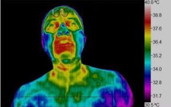
Rainbow images (heat patterns). A colour scale mapping temperature variation — warmer areas in reds and oranges, cooler areas in blues and greens. These make it easy to see where blood flow is higher or lower.
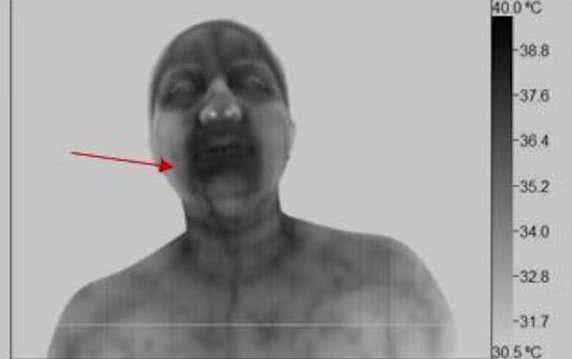
Inverse grayscale images (vascular patterns). These reverse black and white tones to emphasise blood vessel activity and circulation, complementing the heat maps and giving a fuller picture.
Independent expert interpretation. Your images are not read by the technician who takes them. A board-certified thermologist reviews every study, explains what the patterns indicate, and notes anything that warrants follow-up with your healthcare provider.
Who Interprets Your Images
Robert L. Kane, DC, DABCT, FIACT has worked in thermal imaging since 1992. He holds Diplomate certification in Clinical Thermography from both the American Board of Clinical Thermography and the International Academy of Clinical Thermology, which named him a Fellow for his contributions to the field. He studied breast applications under Dr. William Hobbins, one of the field’s foremost authorities. He has served on the boards of both IACT and ABCT, trains thermography technicians, and has lectured on breast thermology at the Cleveland Clinic, Cedars-Sinai and the American Academy of Thermology. He runs an interpretation practice serving clients across the United States, Canada, Israel and the Philippines.
What to Expect During the Procedure
A non-invasive, gentle process. An infrared camera captures your body’s natural heat emissions. No radiation, no compression, no contact of any kind. The camera never touches you.
Region-by-region imaging. You will stand or sit in front of the camera while each area is imaged. Each region takes a few minutes. Before imaging begins we will ask you to spend 10–15 minutes in the room so your skin temperature can settle — this step is what makes the underlying patterns readable.
A private, comfortable setting. Imaging takes place in a temperature-controlled private room. Our technician will guide you through positioning with care.
Nothing to feel. No noise, no discomfort, no need to hold still for long stretches.
Afterwards. No recovery time — you are free to go as soon as imaging is finished. Your report follows within 7 business days.
How to Prepare for Your Appointment
These steps matter more than they might seem. Skin temperature is what the camera reads, so anything that heats, cools or restricts circulation beforehand can obscure the patterns we are trying to see.
Avoid activities that affect temperature or circulation
- For at least 24 hours prior, avoid sun exposure or sunburn to any area being imaged
- No massage, chiropractic care, acupuncture, physical therapy, ultrasound, dry needling or laser therapy on or near the areas being imaged
- No ice packs, heating pads, saunas, TENS units or electrical muscle stimulation within 24 hours prior
- No strenuous exercise for 4 hours beforehand
- No smoking, alcohol or caffeine for 4 hours beforehand
Dress comfortably
Wear loose clothing that is easy to remove or adjust. Avoid tight garments that restrict circulation or leave marks on the skin. If you wear external breast prostheses, remove them at least 12 hours prior.
Hair
Please bring a hair tie. Hair must be worn up and away from the face, not covering the forehead, neck or shoulders.
Keep skin clean and product-free
- No lotions, oils, creams, powders or makeup on any area being imaged
- No deodorant or antiperspirant
- Do not shave any area being imaged for 24 hours prior
Before your appointment
- Do not bathe or shower within 1 hour of your scan
- If medically appropriate, avoid pain medication or vasoactive drugs beforehand — check with your doctor if you are unsure
- If you are nursing, do not nurse within 1 hour of the exam, and allow as much time as you can
Health considerations
Recent sunburn or skin burn may mean we need to reschedule. Please bring any prior imaging reports or documentation relating to areas of concern.